By Frangiska Mylona,
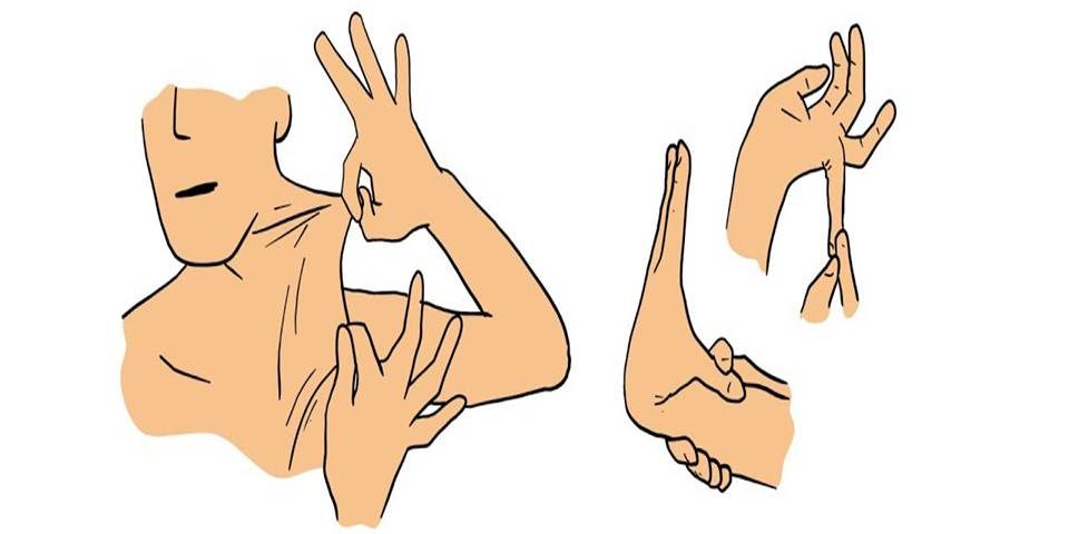
Ehlers-Danlos syndrome (EDS) is a rare genetic, heterogeneous group of connective tissue disorders caused by mutations in genes responsible for collagen synthesis which is the main component of connective tissue. The EDS is divided into thirteen types with different manifestations and variable ways of inheritance. Two of its main symptoms are hyperelastic skin and hyperflexibility of joints.
In order to understand the pathophysiology of this syndrome, it is necessary to dig into the microscopical world of human tissue architecture and especially in the connective’s tissue structure.
Specifically, the human body consists of four major types of tissues:
- Epithelial tissue; covers the body and its organs
- Connective tissue; binds tissues together and supports them
- Muscle tissue
- Nervous tissue.
In general, the connective tissue consists of cells and an extracellular matrix which is the non-cellular portion of the tissue providing a physical scaffolding for the cellular constituents and has the shape of a complex mesh-work made from protein fibers like collagen, elastic and reticular fibers as well as an amorphous component of carbohydrates and proteins that forms the tissue’s ground substance providing its gel-like appearance. The most fascinating aspect of this type of tissue is that it forms a vast and continuous compartment throughout the body while being bound to the epithelial tissue by the basal lamina which acts as a base anchoring the connective tissue to the various epithelial types.
Apart from being anchored to the epithelial tissue by the basement membrane and providing the body’s structural framework, it helps in the protection of delicate organs and in the interconnection between various tissue types. In addition, it aids in fluid transportation between the tissues and it acts as a line of defense against microorganisms.
Connective tissue encompasses many types with different functional properties but with many common characteristics. The types of connective tissue are listed as follows:
- Embryonic connective tissue includes the mesenchyme and the mucous connective tissue
- Connective tissue proper includes the loose connective tissue and the dense connective tissue which is further subdivided into regular, which is the major component of tendons, ligaments, and aponeuroses, and the dense irregular connective tissue including the reticular connective tissues found in the dermis of the skin
- The specialized connective tissue includes cartilage, bones, adipose (fat) tissue, blood, hematopoietic tissue, and lymphatic tissue.

As it was mentioned above, Ehlers-Danlos syndrome is caused by mutations in genes encoding collagen’s structure, where collagen plays an active role in the extracellular matrix of the connective tissue. There are many collagen types (around twenty-nine in number) with the most well-known and studied being the following:
- Collagen type 1; is found in skin, bone, tendons, ligaments, organ capsules, and others providing resistance to force, tension, and stretch.
- Collagen type 2; is located in the intervertebral disks and in the cartilage, providing resistance to pressure.
- Collagen type 3; is found in loose connective tissue and organs such as the uterus, liver, spleen, kidneys, lungs, blood vessels, fetal skin, and others. It functions by providing a supportive meshwork for the organs and the blood vessels.
- Collagen type 4; is found in the basal lamina of the epithelial cells, kidneys, and in the lens providing support and a filtration barrier.
- Collagen type 5; is distributed uniformly throughout the connective tissue stroma. The mutations that lead to the abnormal formation of collagen involve many genes located in different chromosomes. Indicatively, some of them are:
- The COL1A1 gene is located on the 17th chromosome and is responsible for the production of type 1 collagen
- The COL1A2 gene located on the 7th chromosome is responsible for the production of collagen types 1 and 2
- The COL3A1 gene on the 2nd chromosome is responsible for producing collagen type 3
- There are two genes, COL5A1 located on the 9th chromosome, and COL5A2, located on the 2nd chromosome, both producing collagen type 5
- Others like ADAMTS2,TNXB,PLOD1, etc.
A total of thirteen types of Ehlers-Danlos syndromes are classified and some of them are very rare. In this article, only eight of its types will be analyzed. About the:
- EDS type I or Classical EDS (cEDS) is characterized by extremely thin, elastic, and velvety skin that is easily injured. Also, other symptoms that are observed are hyperflexible joints, abnormal scarring leaving atrophic scars, varicose veins, and aortic rupture. It is inherited in an autosomal dominant pattern, so there is a 50% chance that a child will inherit it from an affected parent. The responsible mutated genes are COL5A1, COL5A2, and most rarely, COL1A1.
- EDS type II or Classical-like EDS (clEDS) is characterized by; milder symptoms than EDS type I with the absence of atrophic scars. As it is inherited in an autosomal recessive manner, even if the parents are healthy while carrying the mutation, they still have a 25% chance of passing this mutation on to their children. The responsible mutated gene is the TNXB.
- EDS type III or hypermobile EDS (hEDS ) is characterized by; asymptomatic joint hypermobility. The genetic cause of hEDS has not yet been identified but it is inherited via autosomal dominant inheritance. A variety of conditions can accompany hEDS, although not enough evidence exists for them to become diagnostic criteria because they are not highly specific for EDS and there is no proof they are the result of hEDS. These symptoms are; sleep disturbance, fatigue, postural orthostatic tachycardia, functional gastrointestinal disorders, anxiety, and depression.
- EDS type IV or vascular EDS (VEDS) is characterized by; aortic rupture at a young age, very fragile and elastic skin, easy bruising, and other severe symptoms. The mutated gene that seems to be responsible is COL3A1, inherited in an autosomal dominant manner.
- EDS type VI or kyphoscoliotic type is characterized by; congenital muscle hypotonia with early kyphoscoliosis and dislocations in the joints of the shoulders, hips, and knees. PLOD1 is the responsible gene carrying the mutation, and it is inherited in an autosomal recessive manner.
- EDS type VIIA and VIIB or Arthrochalasia EDS (aEDS) is characterized by; congenital, bilateral hip dislocation and hyper-extensible skin. The COL1A1 and COL1A2 mutated genes seem to be responsible for the disease which is inherited as an autosomal dominant trait.
- EDS type VIII or Periodontal EDS (pEDS) is characterized by additional symptoms which may include; severe periodontitis at an early stage, tooth loss, skin irregularities, and lack of attached gingiva. Mutations in the C1R gene appear to be the cause for this type of EDS where normally it plays a role in the activation of the complement system that is part of the immune system. It is inherited in an autosomal dominant manner.
- Cardio-valvular EDS (cvEDS) can cause serious cardia-valvular problems such as mitral valve prolapse, hyperextended and thin skin that is easily bruised, forming atrophic scars, and abnormal joint movement. The responsible mutated gene is COL1A2 and the disease is transmitted in an autosomal recessive pattern.
Aside from the autosomal dominant and autosomal recessive types of inheritance, EDS type IX is inherited in an X-linked recessive manner. As a result, biological males have a higher chance to be affected by this type of EDS, while biological females have a lower risk of being affected and are frequently carriers of the disease.
A diagnosis of EDS is made based on a combination of Beighton and Brighton criteria, which give a score based on the symptoms and clinical image of the patient. Subsequently, both of their scores are added and we can categorize and assess the patient.

The Beighton score focuses on generalized joint hypermobility on a 9-point scale that involves the joints of:
- The fifth finger. For example, both sides are tested by resting the palm and the forearm on a flat surface with the palm side down and fingers out straight. If the fifth finger is bent upwards and extended backward beyond 90 degrees, one point will be added for each hand.
- The base of both thumbs
- Elbows
- Knees
- Spine.
The angle that is formed is calculated by a goniometer.
The Brighton criteria are divided into:
- Major criteria involving arthralgia (pain in the joints) for more than three months in four or more joints while also including the Beighton score that it must be above or equal to four
- Minor criteria; including the Beighton score of 1, 2, or 3, the dislocation of more than one joint, skin hyperextensibility, the number of skin lesions, the Marfanoid habitus of the patient that is characteristic of the collagen type of diseases like if the patient is tall, slim with arachnodactyly, the presence of varicose veins or, the detection of mitral valve prolapse, etc.
The patient must have a combination of the following;
- Two major criteria
- One major and two minor criteria
- Four minor criteria
- Two minor criteria and a first-degree relative who has been diagnosed with EDS. Additionally, the diagnosis should include molecular analysis to detect mutations in genes involved in collagen formation or other genes associated with the EDS.
Aside from supportive care, there is no definitive treatment for EDS complications and pain. The therapeutic approach includes surgical interventions for aortic rupture or mitral valve prolapse, physiotherapy, and drugs with analgesic properties.
In conclusion, although patients with EDS might feel a restriction regarding their mobility, they can live normal lives. However, because they are prone to fatal complications like aneurysms due to the fragile blood vessels, early diagnosis, and treatment can be lifesaving, especially when it comes to disorders that have so many types and such a broad spectrum of symptoms.
References
- Histology; A Text and Atlas with correlated cell and molecular biology by Wojciech Pawlina.Seventh Edition.
- Genomic Medicine ,2016, Sofia , Prof. Draga Toncheva , Prof. Varban Ganev.


