

By Frangiska Mylona,
There is a common saying stating that sleep and death are “first cousins” or even “twin brothers” in comparison to the states that people are in when they fall asleep and when they die. Throughout history, people have been fascinated by the fact that the body cannot voluntarily control its actions when sleeping and dying, and for this reason, both situations have been compared.
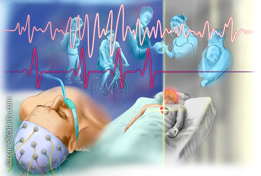
Ancient Egyptian culture depicted this connection between the afterworld (the netherworld for the ancient Egyptians) and the world of dreams, as well as the ingrained relationship between sleep and death along with resurrection and awakening. Furthermore, ancient Greeks believed Nyx (“Night”) and Erebos (“Darkness”) had twin sons called Thanatos (“Death”) and Hypnos (“Sleep”). Using a medical procedure known as an electroencephalogram (EEG), it is possible to visualize the connection between these two states. As a result, death does not appear suddenly by “shutting down” the brain but rather it can produce an electrical activity similar to the electrical activity observed in sleep.
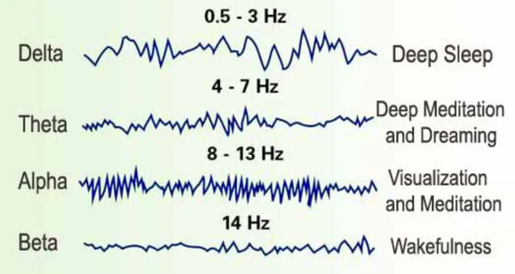
Electroencephalogram (EEG) records the electrical activity of the cerebral cortex via electrodes placed on the skull. The EEG waves are not action potentials, as commonly mistaken, instead, they originate from alternating excitatory and inhibitory synaptic potentials that produce an extracellular current flow which is detected by the surface electrodes. The normal EEG consists of waves that present with different frequencies and amplitudes regarding the state of the patient. For instance, when the adult patient is awake with their eyes open beta-waves (13-30 Hz) are observed. If the person is actively engaged in solving a problem or deeply focused, gamma waves (30-100Hz) will be produced. On the other hand, if the patient closes their eyes, alpha waves (8-13Hz) are detected.
According to some sources, there are four stages of sleep, while others claim that there are five stages. Nevertheless, these stages fall into two categories, the Non-REM (NREM or Non-rapid eye movement) sleep, which occurs first and includes three or four stages, and the REM (rapid-eye movement) sleep, during which dreams are usually experienced. NREM stage N1 is the transition of wakefulness to sleep that lasts for a few minutes and it is the lightest stage of sleep. In this staged alpha-waves are intermixed with low-frequency theta-waves (4 to 7 Hz). The muscles begin to relax and the heart starts to slow down along with the breathing rate. NREM stage N2 is the stage before the person enters into deep sleep. There are no eye movements and both heart and breathing rates along with body temperature start to drop. In this stage, EEG detects low-frequency waves with sleep spindles (high-frequency bursts) and K complexes (large, slow potentials).
NREM stage N3 signals the deepest sleep stage where the arousal from sleep is difficult whereas the body is fully relaxed since the heart beats along with the breathing rate, are at their lowest levels. At this stage, it is observed a renewal of the organism since cell regeneration and tissue repair occur as well and the immune system gets reinforced. At that stage, EEG detects delta waves with low frequency along with occasional sleep spindles. It is followed by NREM stage N4 which is characterized by delta waves and lasts for almost 30 minutes. Around every 90 minutes, the slow-wave sleep pattern changes to rapid eye movement (REM) sleep. The EEG waves become similar to when the person is awake since it detects desynchronized high frequency with low voltage waves like beta waves (14-30Hz) and gamma waves (30-100Hz).
This similarity leads to the REM stage being known as ‘’paradoxical sleep’’ since the morphology of the waves resembles the ones observed during wakefulness however the person is in the state where it is the most difficult to get awaken. The REM stages can also be divided into two phases; the phasic and tonic where only in the phasic phase, rapid eye movements are observed. Generally, in this stage, dreaming occurs along with the increase in breathing and heart rates. Another interesting phenomenon is observed, known as muscle paralysis with occasional twitching. This twitching is known as the “Hypnic Jerk” and is more likely to occur if the person is under a lot of stress, coffee, stimulants, etc… The proportion between the NREM state and the REM state varies in the human lifespan with the REM state presenting a decline in its duration as the person gets older.
In this article, two studies will be analysed regarding the results obtained from the brain activity of dying patients. The first study was conducted by Frontiers in Aging Neuroscience where they analysed the EEG of a dying 87-year-old male patient. The patient experienced a heart attack after a subdural hematoma. He also underwent a craniotomy for the hematoma to be removed. On the EEG of the 87-year-old male patient, four-time points were examined, including the seconds after the clinical seizure, the suppression of left and bilateral hemisphere activity, and the phase of cardiac arrest until the end of the recording. The conclusions were as follows. Throughout the entire EEG recording, there was exhibited spectral activity levels below 25 Hz. A reduction in delta brainwaves was observed between the clinical seizure and after the suppression of the left hemisphere. At the time of cardiac arrest, a little increase in the delta waves was observed, although they ultimately declined at the end of the EEG recording.
At the time between the suppression of the left and bilateral hemispheres, absolute theta activity decreased and remained stable throughout the period between the cardiac arrest and the end of the EEG recording.
After bilateral hemispheric activity ended, there was a temporary increase in gamma brainwaves but these levels quickly declined after the patient experienced cardiac arrest. It is imperative to notice that gamma waves are also associated with memory therefore there is an assumption that during this time, the brain may perform a memory flashback.
Another study was conducted in the Loyola University Medical Center, May-Wood III from January 1984 through May 1986. Fifty-six consecutive patients were clinically diagnosed as brain dead. Interestingly, eleven of them had electroencephalographic (EEG) activity following the diagnosis of brain death. Even though the EEG showed brain activity, none of the patients recovered. The mean duration of the observed EEG activity was 36.6 hours and three EEG patterns were observed;
1) 9 patients were observed with theta-/ or beta-waves of 72 hours following brain death.
2) 2 patients were observed with mixed sleep-like activity with delta-/theta- waves and spindle-shaped patterns 168 hours after brain death.
3) 1 patient was observed with alpha-activity 3 hours following brain death.
One of the conclusions of this study was to question the use of EEG as a confirmatory test of brain death. Yet, it is incredible to notice that on the verge of dying and even after brain death is announced, some patients exhibit brain electrical activity similar to the state of the brain’s electrical activity during sleep!
It is crucial to keep in mind that several factors can contribute to the observed EEG activity during the time of death, including the conditions that the patient is in like oxygen availability, the cause of death as well as the impact of anesthesia on the patient.
As a conclusion, the similarity between brain activity during death and sleep is intriguing, as is the transition of human brain function during death as well as after death. It is a comforting realization that death does not just “turn off the brain” but it leads the body into a deep, peaceful state similar to sleep or even dreaming. These conclusions can soothe the hearts of those left behind after the death of their loved ones. Perhaps, death is not as cruel as it is thought to be, maybe death is not a state of complete darkness and immobility, but the entrance to new dimensions, unimaginable to those who are still living, yet as peaceful as sleeping.
References
- Physiology 6th Edition by Linda S.Constanzo Ph.D.
- Researchers study EEG patterns in dying men to understand brain activity at the end of life. Available here
- Electroencephalographic Activity After Brain Death. Available here
- Internet articles; Everything to Know About the Stages of Sleep. Available here