

By Fanis Michalakis,
We have all heard or seen bioluminescence since we all know about fireflies or angler fish from movies or videos. This phenomenon has fascinated humans for centuries with the earliest reference in ancient Chinese texts. In ancient Greece, Aristotle would describe the light observed on some animals. Similar references would be made in the next years. Later in the 19th century, the experiments of Raphaël Dubois led him to the scientific discovery of the mechanism.
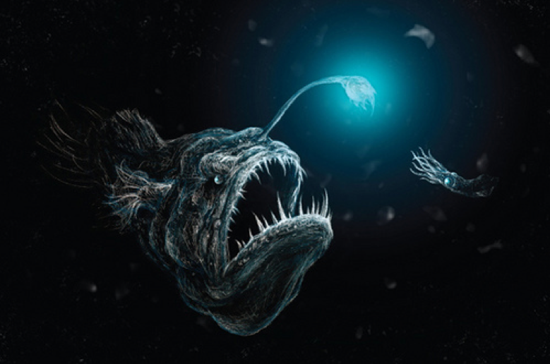
First off, the light of bioluminescence is produced when a substance called luciferin reacts with an enzyme called luciferase with the requirement that there is oxygen around. Bacteria, fish, insects, and squid have been observed using bioluminescence. However, between two different groups and even between species the two key components can have differences but the result is the same. Therefore, the firefly and the squid use different molecules with different properties, each adapted best to their needs. The light produced can be a way of communication between two animals as is the case with the fireflies or it can be used to attract prey closer, a method used by the angler fish, which uses bacteria that are inside its esca (bait) to draw its prey in front of its mouth.
Another function that the light can serve is defense, in the sense that either the predator is startled, the prey will glow in the stomach of the predator, and then a transparent predator can be easily spotted by others, or the light draws bigger predators to its location. Moreover, the light can help will illuminant the darkness of the ocean or counterillumination, which is a way for animals to cloak their bodies in the sea. Basically, they produce the same dim light that comes from the surface which hides the shadow that they would otherwise produce. Some researchers have suggested that bioluminescence can also be a way to warn predators about venom on the body of its prey or just a by-product of its metabolism.
As is the case with any new characteristics that we discover, we have tried to use bioluminescence imaging in our everyday lives. More specifically, we have tried to detect pathogens in an animal’s body through this technology. Traditionally, to study a pathogen, its infection, its stages, the development rate, and the interactions with the host, researchers infected mice with the pathogen that was of interest and killed and studied them at different time frames. The main idea was that with more time frames we would have more information about the pathogen. However, using bioluminescence imaging, we do not have to kill many animals to analyze the pathogens and we can see the infection happen in vivo, meaning inside a living organism in real time.
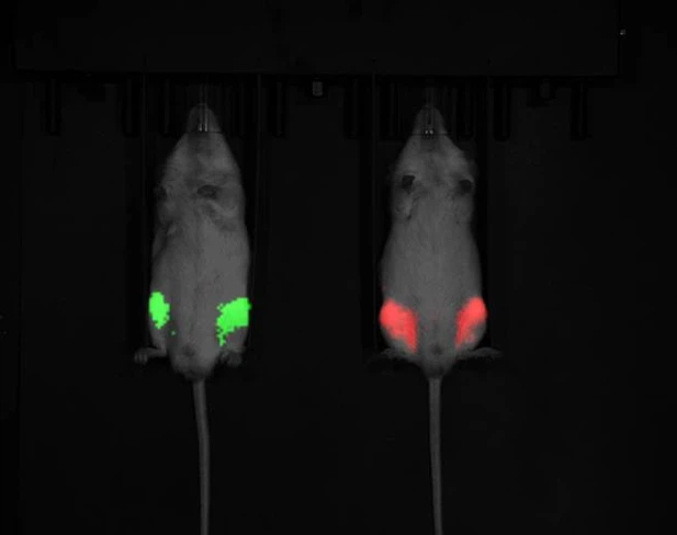
To set up the experiment, we must add the lux operon to a pathogenic microbe and inject it into an animal. The lux operon contains the genes necessary to produce luciferin and the luciferase enzyme. Thus, the pathogens will produce light and we will be able to see the tissues that can be found at the site of the infection. Another way we can set up the experiment is by making the cells of the infected animal or the pathogen able to create the luciferase. In this method, however, we must also add luciferin to the tissues through injections before observation as the host cells cannot produce it themselves. We can then read the emissions produced on the sites of infection.
This experiment has already been done on mice with promising results. It should be noted that the method has its benefits and its shortcomings. It allows us to detect the pathogens in all the tissue samples with ease, whereas other methods could have missed the pathogens in some tissues if the researchers did not think to check those areas. It also allows us to detect pathogens even if the colonies are small, because of this method’s small background signal. Apart from microbes, we can observe how a bigger parasite interacts with its host and the different stages of its life cycle by making the parasite produce light only at certain stages.
However, despite its benefits, this method will probably not be used on humans. Every organ has a pigmentation that can influence the reading we receive. The alteration becomes more extreme in deeper tissues as there are more layers of organs on top. Since humans have more layers of tissue in their bodies, this method would not work for us. It is also hard to detect two infection sites that are close to each other. Moreover, to study the organism, we must first apply some changes to make it glow. This process can be more difficult for some organisms and as such, this technology might not be suitable for them. Lastly, we need oxygen in the area for the reaction to happen, so some tissues like the intestines will yield negative results.
In conclusion, bioluminescence is more than a glowstick that animals flail around aimlessly. Although in our minds we might think that all of it is the same, different animals use different mechanisms to achieve the same result but for different reasons. Due to its shining nature, apart from wild animals, this trait has also drawn our attention, leading to many experiments.
References
- The history of Luciferin and Luciferase. goldbio.com. Available here
- Applications of bioluminescence in biotechnology and beyond. pubs.rsc.org. Available here
- Bioluminescence in the Sea. digitalcommons.calpoly.edu. Available here
- Bioluminescence. ocean.si.edu. Available here